- Price: 1$
- Size Facebook: 1702 x 630 px
- Size Twitter: 1500 x 500 px
- Size LinkedIn: 1128 x 191 px
More Facebook, Twitter and LinkedIn Cover Photos
Cover photo info
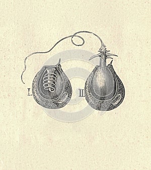
- Photo title: Antique engraved illustration of the hydra stinging fibre. Vintage illustration of the hydra stinging fibre. Old
- Author: Romannerud
- Cover photo description:
- Antique engraved illustration of the hydra stinging fibre. Vintage illustration of the hydra stinging fibre. Old engraved picture. Longitudinal cross section of the hydra. Book illustration published 1907. Hydra has two main body layers, which makes it diploblastic . The layers are separated by mesoglea, a gel-like substance. The outer layer is the epidermis, and the inner layer is called the gastrodermis, because it lines the stomach. The cells making up these two body layers are relatively simple. Hydramacin is a bactericide recently discovered in Hydra it protects the outer layer against infection. A single Hydra is composed of 50,000 to 100,000 cells which consist of three specific stem cell populations that will create many different cell types. These stem cells will continually renew themselves in the body column. Hydras have two significant structures on their body: the head and the foot . When a Hydra is cut in half, each half will regenerate and form into a small Hydra the head will regenerate a foot and the foot will regenerate a head . If the Hydra is sliced into many segments then the middle slices will form both a head and a foot . Respiration and excretion occur by diffusion throughout the surface of the epidermis, while larger excreta are discharged through the mouth.
- Image ID:262181566
- Views:0
- Downloads:0
Keywords for Facebook, Twitter and LinkedIn timeline photos
wildlife
vintage
picture
old
natural
image
illustration
history
historic
heritage
graphic
fauna
engraving
engraved
drawing
culture
cultural
century
book
artwork
art
archive
archival
antiquity
antique
animal
ancient
aged
age
19th
1800s
1800
zoology
hydroid
structure
hydrozoa
cnidaria


